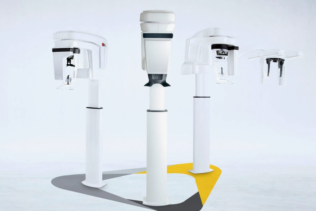
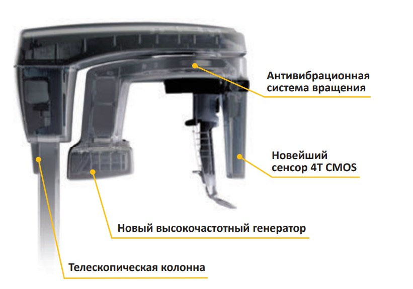
Physicians and specialists can perform 2D and 3D imaging with one compact system available at a reasonable price. The CS 8100 3D system from Carestream Dental allows doctors to perform a greater number of different procedures. It incorporates the latest 4T CMOS sensor technology, which reduces imaging noise and provides detailed contrast images at low radiation doses. The versatility of the system lies in the ability to perform two-dimensional panoramic examination, three-dimensional “tomographic” imaging and scanning of three-dimensional models.
In addition, the device itself is very compact and meets the latest ergonomic requirements. The CS 8100 3D incorporates panoramic 2D imaging technology that provides overview images in seconds. A set of diagnostic programs simplifies a range of examinations, including panoramic (for adults and children), segmented panoramic imaging, temporomandibular joint imaging, and maxillary sinus imaging. The greater thickness and width of the focal area provides a greater margin of error for patient positioning in complex anatomical cases.
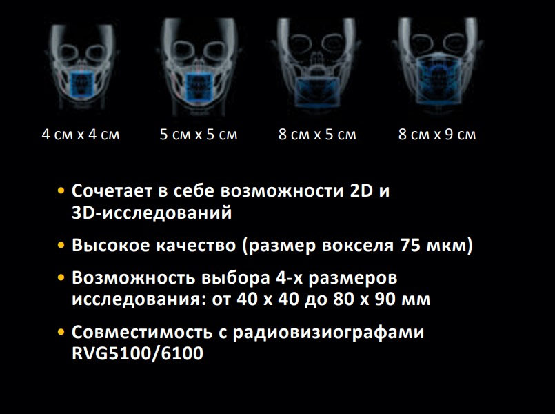
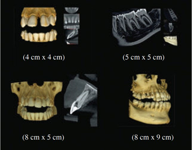
The CS 8100 3D system incorporates three-dimensional imaging technology that allows for 1:1 images without distortion or overlapping of anatomical elements. A choice of programs gives physicians the ability to set the image size and level of detail, as well as the radiation dose for each individual case. These programs provide a universal viewing area (5 cm x 5 cm) for everyday dental examinations such as local pathology, single implants and endodontic examinations. It is possible to scan either one (8 cm x 5 cm) or both (8 cm x 9 cm) jaws and dental arches. The special program (4 cm x 4 cm) is ideal for pediatric examinations, while the EndoHD mode (5 cm x 5 cm) provides a high-resolution three-dimensional examination with 75 µm grain size and captures the smallest details of root and canal morphology. Professionals can also create highly accurate 3D models with the CS 8100 3D system by scanning traditional casts, X-ray templates and plaster models. The resulting data can be integrated into a computer-aided design system for use with CAD/ CAM systems.
All of these diagnostic capabilities are combined with an intuitive user interface and a computerized system in which only four operations are required to obtain an image: select a program, place the patient in a given position, take the image and analyze it. The CS 8100 3D bite block and positioning system simplifies correct patient positioning and reduces the risk of having to reshoot.
The CS 8100 3D system features motorized height adjusters and is suitable for patients of all heights and clients in wheelchairs. The device features a vibration-free rotation system and rigid locking devices with handles to assist patients through the examination. Research is simplified by the fact that images are obtained very quickly. In Flash Scan mode, the entire procedure takes only seven seconds. This speed minimizes both the duration of x-ray exposure and the risk of patient displacement.
With comprehensive imaging software, physicians can analyze images, use implant placement planning features, and show images to patients so they can better understand the proposed treatment plan. X-ray images can also be transferred to portable USB flash drives, burned to CDs and DVDs, e-mailed and screen shots can be taken. To enable easier collaboration on individual cases, the full-featured 2D and 3D image viewing software can be shared with colleagues and dentists without restriction.

