
MARINA CHIBISOVA,
Doctor of Medical Sciences, Professor of the Department of Clinical Dentistry, Professor of the Y. A. Fedorov Chair of Pediatric and Therapeutic Dentistry of the Federal State Budgetary Educational Institution of Higher Education “I.I. Mechnikov North-Western State Medical University”, Ministry of Health of the Russian Federation, Professor of the Department of Therapeutic Dentistry of the Federal State Budgetary Educational Institution of Higher Education St. Petersburg State University
MODERN 2D- AND 3D-DIAGNOSTICS
Radiologic diagnostic methods are now widely used in therapeutic, surgical, orthopedic, pediatric dentistry and in orthodontics. Radiography is one of the most important diagnostic methods, giving the most complete picture of the structure of teeth and surrounding tissues, adjacent anatomical areas, the study of the state of which is necessary for the complex treatment of patients with various clinical forms of dental diseases. Intraoral dental radiography and panoramic zonography of the dento-mandibular system (orthopantomography) are widely used in clinical practice. Currently, new methods of radiation diagnostics are developed and introduced into clinical practice: ultrasound scanning (U/SCBCTCBCT), cone-beam computerized tomography (CBCT), multislice computed tomography (MSCT), magnetic resonance imaging (MRI), etc.
Sophisticated specialized dental radiology complexes for computerized dental radiography (radiovisiography) used in outpatient dentistry are being created.
Radiologic diagnostics in outpatient dentistry, maxillofacial surgery and otorhinolaryngology helps to improve the quality of treatment of diseases of the dento-mandibular system and maxillofacial region
Digital panoramic radiography (orthopantomography), cone-beam computed tomography (CBCT) and dental radiovisiography are becoming the standard for patient examination when drawing up a dental treatment plan and are used for dynamic monitoring in any section of the outpatient practice of dentists. 2D (OPTG, TRG, radiovisiography) and 3D (CBCT) digital radiodiagnostics sufficiently ensures radiation safety for patients and medical personnel during examinations.
Processing of the X-ray image by appropriate computer programs makes it possible to create, store and update the data archive of electronic medical records of patients.
Differential diagnosis becomes more rapid and reliable. It becomes possible to store and analyze all necessary information on diagnosis and treatment of diseases and injuries of teeth, jaws, temporomandibular joints, maxillary sinuses.

Modern dentistry represents one of the most rapidly progressing branches of medicine. Successes in the treatment and rehabilitation of patients with disorders of the dento-mandibular system are due to the wide use of unique science-intensive technologies, the latest specialized materials, equipment, facilities and instruments that meet the highest requirements. This is equally true for radial diagnostics in dentistry, as the efficiency and results of treatment significantly depend on its level.
Radiologic examination serves as one of the leading methods for diagnosing most diseases and injuries of the dento-mandibular system, as well as the maxillofacial region in patients of different age groups. Radiographic examination is necessary to clarify the diagnosis, to determine the plan and prognosis of treatment, to study the changes occurring in patients of different age groups, as well as under the influence of therapeutic measures.
Depending on the purpose, it is important to choose the most effective method of radiologic examination.
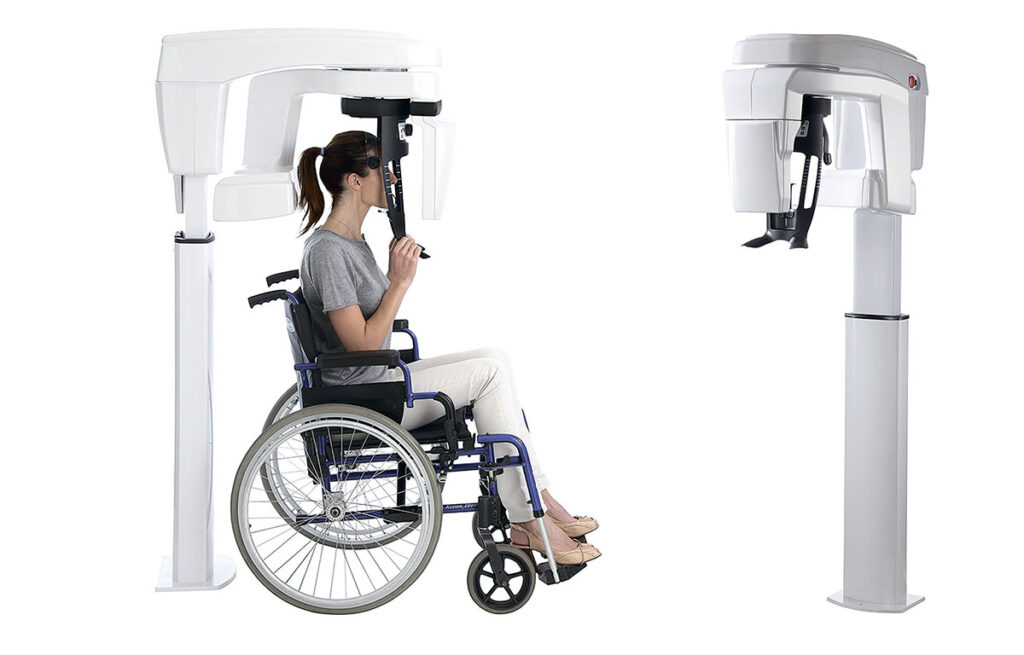
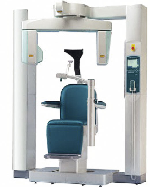
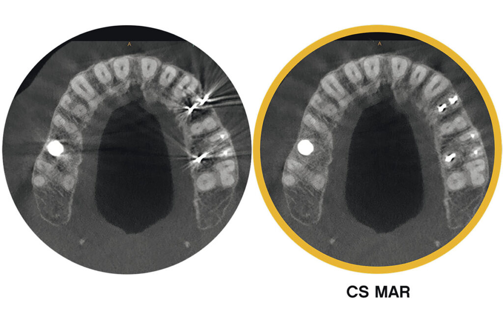
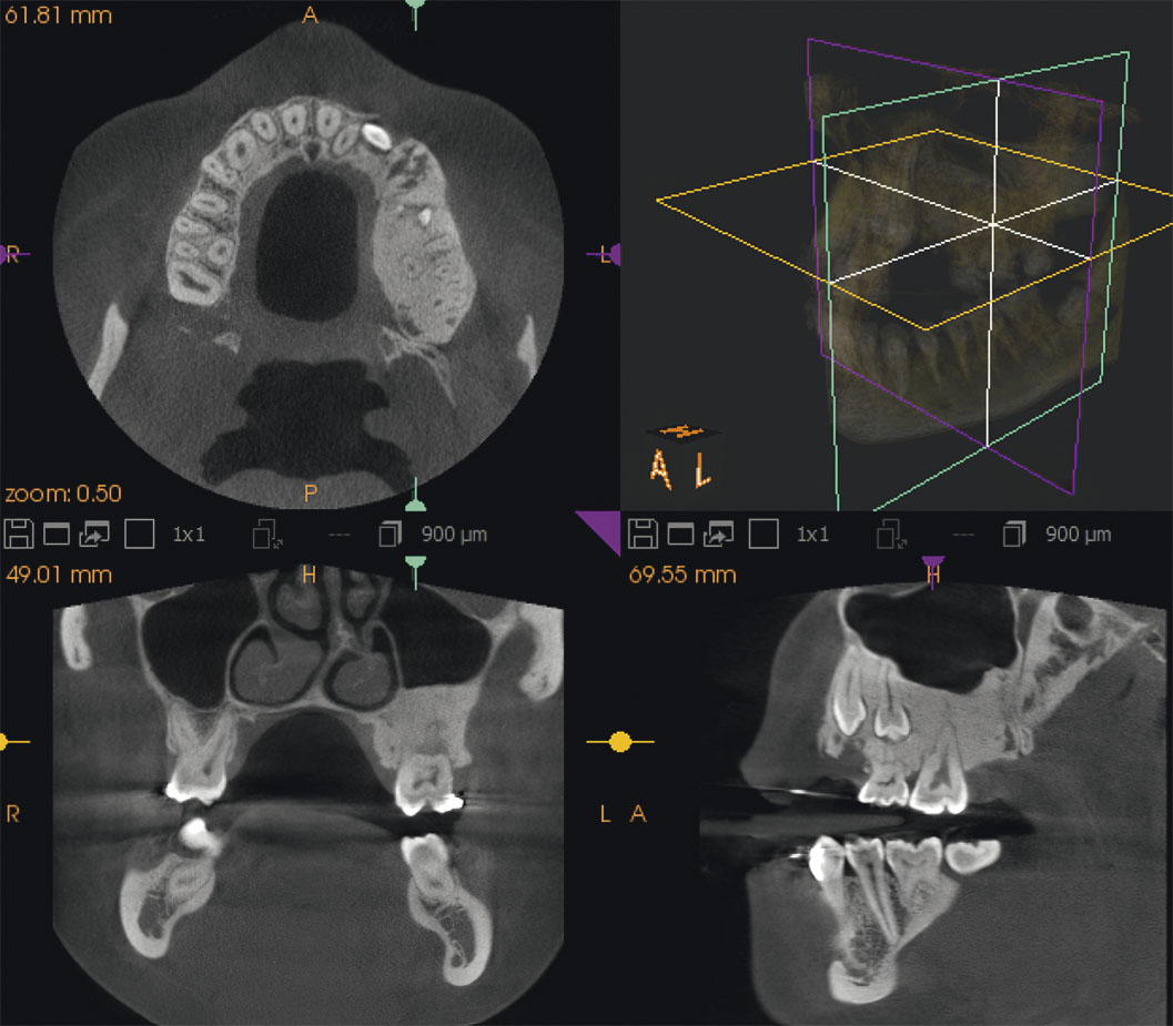
Intraoral radiovisiography, orthopantomography (OPTG), and cone-beam computerized tomography (CBCT) are the most widely used (Fig. 1; Fig. 2 (A, B)).
In outpatient dentistry, all radiological examinations of patients (mandatory and additional) are prescribed by the dentist, justifying the necessity of radiological diagnostic methods associated with the use of ionizing radiation sources from a clinical point of view.
Currently, many private and public outpatient dental clinics have dental three-dimensional computed tomography scanners on which 2D and 3D examinations (OPTG, TRG, CBCT) are performed. In addition, according to modern regulatory requirements (Order of the Ministry of Health of the Russian Federation No. 786) it is allowed to place radiovisiographs not only in a separate office, but also in the dental office near the chair. Dental CT scanners are also installed in hospitals where outpatient and inpatient treatment of patients in maxillofacial surgery and otorhinolaryngology is performed. As part of the compulsory health insurance, the patient is offered dental radiovisiography to draw up an examination plan and assess the results of treatment of dental caries and its complications. All other extra-oral radiologic examinations are performed as part of paid medical services (OPTG, TRG, CBCT)
The CS 8100 3d dental 3D tomography scanner complies with all EEC and international medical standards.
The first CBCT machine from J. Morita (Japan) was installed in a private dental clinic in St. Petersburg in 2006
Non-profit professional organizations СтАР (Stomatological Association of Russia, section “Radiation diagnostics in dentistry”) and РОРР (Russian Society of Radiologists and Radiologists) have developed Standards for equipping dental clinics with X-ray equipment. Every multidisciplinary dental clinic should have a dental three-dimensional tomograph performing OPTG, TRG and CBCT, and radiovisiographs are placed in dental offices to monitor the quality of dental treatment in all sections of outpatient practice. There is no such thing as a perfect CBCT; each device has its own advantages and limitations.

CS 8100 3D DENTAL THREE-DIMENSIONAL TOMOGRAPH
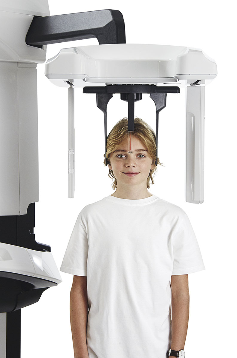
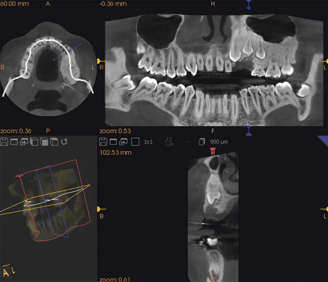
Many modern dental clinics choose the CS 8100 3D from the American company Carestream Dental (Fig. 3). The CS 8100 3D performs lateral, frontal and diagonal X-ray diagnostics.
It consists of the following functional components:
o rotating console;
o shoulder with a control panel;
o panoramic digital sensor;
o x-ray node;
o x-ray control panel;
o chin restraint;
o chin retainer for panoramic photography and bite plate;
o temporal stabilizer; handles;
o nasal positioner;
o software
ADVANTAGES OF CS 8100 3D
Performs high quality 2D (OPTG) and 3D (CLCT) digital images. Carestream Dental also offers fixed and mobile radiovisiographs that can be installed in the dental office. All equipment is certified and has registration permits for use by the Ministry of Health of the Russian Federation.
High-quality physical and technical parameters that comply with European and world standards of radiology.
Low radiation load on patients and medical personnel when performing extra-oral and intra-oral examinations of the dento-mandibular system and maxillofacial region.
Guaranteed service for X-ray equipment.
Convenient, simple software with Russian-language viewer for viewing CBCT data. The viewer can be viewed by dentists in every dental office, it is also possible to record CBCT and 2D images on electronic media and transfer information by e-mail when performing examinations to patients from other clinics.
2D and 3D images are uploaded to the archive for information storage and to the patient’s electronic patient record.
The doses of radiologic examinations can be loaded into the patient’s electronic chart in the dose record sheet.
Optimal cost of the equipment, based on the parameters “price – quality”
Three-dimensional dental computed tomography on cs 8100 type devices has become the basis for planning surgical interventions in inflammatory diseases of the maxillofacial region, implantation and prosthetics of teeth

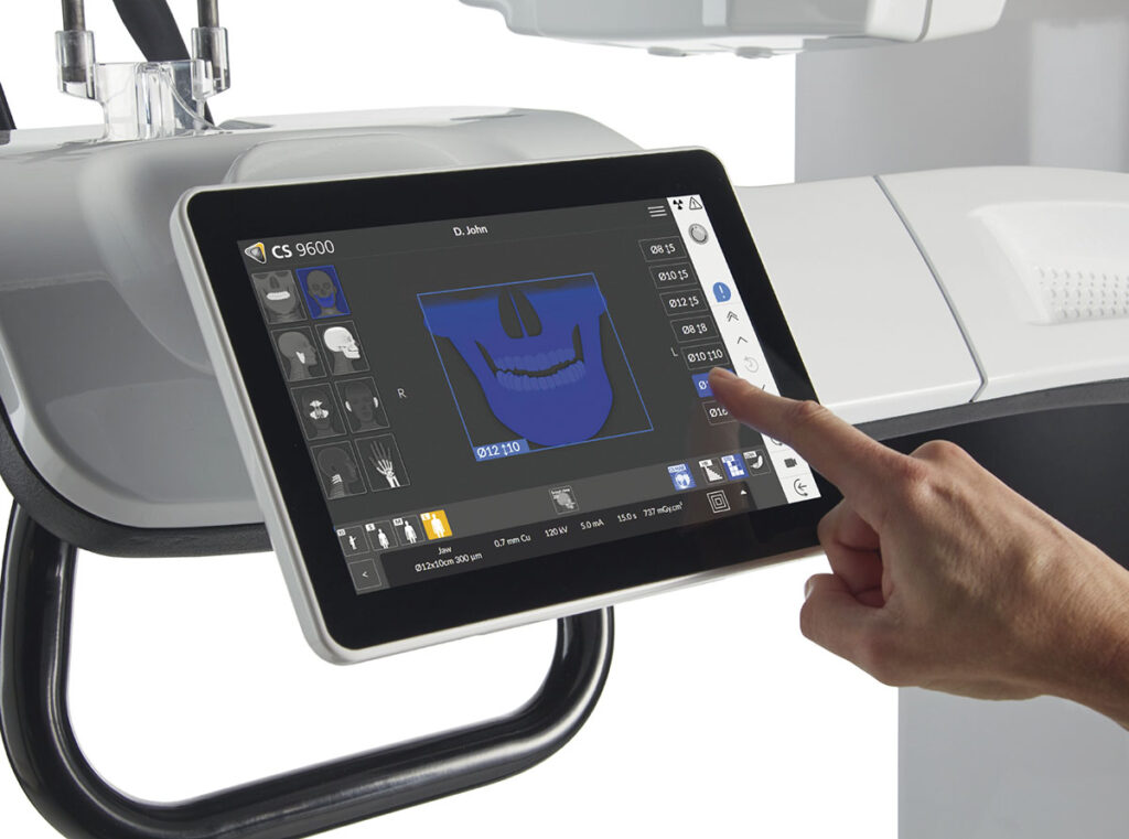
SOFTWARE FOR CS 8100 3D
Dental imaging software is a user-friendly interface that has been designed specifically for x-ray diagnostics. It has the following features:
o has an interface for capturing images;
o ability to manage patient files through the patient window;
o work with intraoral and extraoral images through the image processing window;
o work with three-dimensional images through the window of three-dimensional images processing.
RESULTS OF CS 8100 AND CS 8100 3D IN CLINICAL PRACTICE
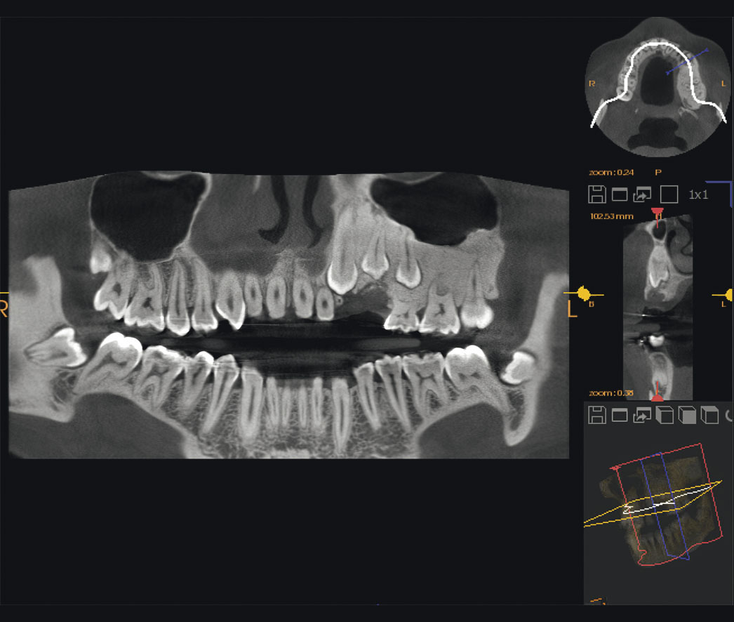
The use of CS 8100 in pediatric dentistry and orthodontics allows to determine the presence of retined teeth, to predict the possibility of correcting their position. In addition, the three-dimensional computer image provides comprehensive information about the state of the jaw bone tissue, which is especially important when selecting supporting teeth at the planning stage of orthodontic treatment. The study of three-dimensional X-ray images allows unmistakable determination of the absence of rudiments, as well as the degree of formation of the crown and root of teeth in children. Computed tomograms reveal the shape, direction and location of the roots of the supporting teeth and the teeth to be moved, clarify the degree of resorption of the roots of primary teeth, the presence and location of the rudiments of permanent teeth, as well as determine retined and supercomplete teeth.
With the help of three-dimensional dental CT it is possible to get a more accurate picture of the degree of mineralization of the roots and crowns of teeth, the degree of resorption of the roots of permanent teeth, the relationship of the roots of permanent teeth with the roots of permanent teeth, the inclination of erupted and retined teeth in relation to adjacent teeth and the median plane.

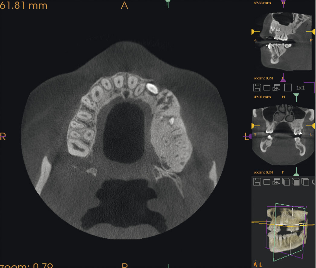
Cone beam computed tomography (CBCT), or three-dimensional dental computed tomography (3D CT), significantly expands the possibilities of radiologic diagnostics, as it provides X-ray images of the structure of teeth and alveolar portion in three projections: frontal, sagittal and transversal. Algorithms of diagnostic examination of dental patients with various pathological processes of the dento-mandibular system, maxillofacial region, facial head, perinasal sinuses and temporomandibular joints are currently largely based on the use of CBCT data.
The experience of using three-dimensional dental computed tomography testifies to the high informativeness of this technique when applied in various sections of outpatient dentistry, maxillofacial surgery and otorhinolaryngology, as well as to the possibility of improving the quality and efficiency of dental treatment on the basis of the obtained data.
The use of three-dimensional dental computed tomography in pediatric dentistry makes it possible to make a correct diagnosis and determine the treatment plan in a timely manner. This avoids complications and reduces the time of examination of children and the radiation burden
To control the state of the tooth after treatment, in dynamics (after 6 and/or 12 months), it is reasonable to perform CBCT, for the reliability of comparative assessment with the initial state. Control panoramic radiography of the whole dentition, or CBCT, is recommended every two years after treatment. CBCT plays an important role in apical surgery, which has now reached a new, higher level in terms of the quality of the procedure – thanks to the use of a microscope and improved methods of retrograde filling of the apical part of the canal after resection of the apical part of the tooth root. Such an algorithm of radiologic examination of patients at the stage of diagnosis, in the course of treatment and during dynamic follow-up of patients in the future allows to obtain the best results of endodontic treatment of teeth, provides reliable methods of prosthetics and other rehabilitation measures for patients (Fig. 6 (A, B, C, D, E)).

